

Plasma-Mediated Femtosecond Laser Ablation
Background : Laser ablation
The use of laser pulses with sub-picosecond durations for the microscopic removal or modification of material has several advantages over the use of longer laser pulses. In the long-pulse regime, material damage is and removal are initiated by a thermal process induced by local heating of material by linear absorption of the long laser pulse. The local heating results in melting, boiling and thermal expansion of the targeted material. Consequently, in this regime, collateral thermal damage can be substantial, and ablation efficiency can exhibit considerable variability due to differences in local absorption within a material and between different material substrates.
Background : Femotsecond Pulse Laser Ablation
Ablation with femtosecond laser pulses proceeds by a distinctly different mechanism than the thermal process observed with longer pulses. With ultrashort pulses, material modification is initiated by a nonlinear absorption process by a pulse that is shorter than the material equilibration time. Femtosecond pulse laser ablation is thus deemed a "non-thermal" ablation process. Although post-pulse equilibration is, of course, still thermal in nature, the use of short pulses with high instantaneous intensity, but low integrated energy, allows for ablation with minimal collateral thermal damage.
The process of material modification with femtosecond pulses begins with a multi-photon absorption that can best be described by as a Zener electron tunneling ionization event. This first seed electron is then accelerated by the large electric field of the laser pulse. An ionization cascade occurs as accelerated free electrons collide with stationary molecules and atoms, producing more free electrons. The cascade proceeds exponentially during the duration of the femtosecond pulse, producing a microscopic neutral plasma, eventually reaching a critical electron density. At this point the high density of free electrons mimics the conduction band in a metal, and the plasma reflects the remainder of laser pulse, and constrains further absorption to a nanometer-scale skin depth within the plasma. All of these dynamics are completed within the ~100 femtosecond duration of the laser pulse, which is shorter than the picosecond-to-nanosecond timescale for thermal relaxation. Thus, all laser-material interactions are strictly confined to the focal volume.
As a result of these self-seeding and self-limiting behaviors, ablation with femtosecond pulses can provide consistent damage across a wide range of materials - from soft tissues to hard dielectric materials to metals, with negligible thermal damage to surrounding structures. Additionally, the highly nonlinear (equivalent to 5th or higher order) initiation process enables material modfication deep to the surface of the target object.
All Optical Histology
 One application of femtosecond pulse laser ablation in my
research has been the invention and development of
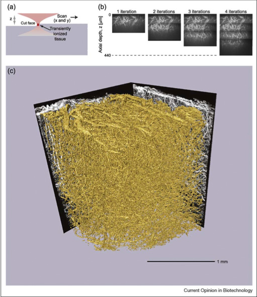
all-optical histology. This technique combines
two-photon laser scanning microscopy and femtosecond pulse
laser ablation to perform serial iterations of imaging and
ablation in histological tissue samples. Here, laser
ablation is used to remove, on a micrometer-by-micrometer
basis, the upper layers of tissue that have previously
been imaged, thereby exposing previously underyling layers
for subsequent round of multiphoton imaging. In
contrast to traditional reconstruction by serial thin
sections, all-optical histology images are taken within an
undisturbed block of tissue, and surface removal of the
previously imaged tissue is performed by non-thermal laser
ablation, which produces no sheer forces upon the soft
tissue block. Thus, we obtain a fully-registered
3-dimensional reconstruction of a macroscopic block of
tissue with micrometer resolution, without the tissue
distortion and mis-registration issues inherent to serial
section reconstruction.
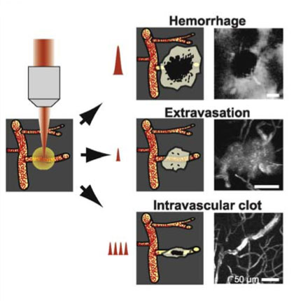
A second application of femtosecond pulse laser ablation has been the invention and development of an in vivo model of stroke. In a research program spearheaded by graduate student Nozomi Nishimura, we utilized femtosecond ablation through a cranial window to produce controlled disruptions to individual capillaries deep within the brain. Depending on the number and energy of pulses, capillaries could be selectively permeated (producing vascular leakage), clotted (producing a microscopic thrombotic stroke), or severed (producing a microscopic hemorrhagic stroke).
A third, currently active, area of research involving femtosecond pulse laser ablation its use in the selective removal of cranial bone. In a research program spearheaded by graduate student Diana Jeong, we are combining femtosecond ablation with optical feedback to produce cranial windows (both traditional craniotomies and PoRTS windows) in rodent skull. The ablation process is continuously monitored to determine the thickness of the remaining bone along with the material composition at the ablation focal volume.